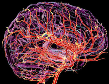
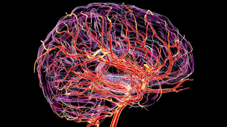
Scientists have visualized the brain’s hidden heartbeat, unlocking new clues to aging and Alzheimer’s.
Scientists at the Mark and Mary Stevens Neuroimaging and Informatics Institute at the Keck School of Medicine of USC have unveiled a breakthrough brain imaging method that captures how the brain’s tiniest blood vessels pulse with every heartbeat. These subtle rhythmic movements may provide new insight into how the brain ages and how diseases such as Alzheimer’s begin to develop.
The study introduces the first noninvasive way to measure “microvascular volumetric pulsatility,” which refers to the rhythmic expansion and contraction of the brain’s smallest blood vessels in living people.
Using an ultra-high field 7T magnetic resonance imaging (MRI) scanner, the team discovered that these microvessel pulses become stronger with age, especially within the brain’s deep white matter—a region vital for communication between different brain networks.
This area is particularly vulnerable as blood flow from the heart’s distal arteries weakens over time. Stronger pulsations in these vessels may disturb brain systems, potentially accelerating memory decline and contributing to Alzheimer’s disease.
“Arterial pulsation is like the brain’s natural pump, helping to move fluids and clear waste,” said Danny JJ Wang, PhD, professor of neurology and radiology. “Our new method allows us to see, for the first time in people, how the volumes of those tiny blood vessels change with aging and vascular risk factors. This opens new avenues for studying brain health, dementia, and small vessel disease.”