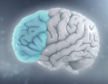
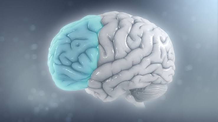
New research shows that brain tissue stiffness helps regulate chemical signals during development. The discovery highlights an unexpected connection between physical forces and the brain’s wiring process.
For decades, scientists have understood that chemical signals, including gradients of signaling molecules, play a central role in directing how tissues grow and organize. More recently, research has revealed that physical factors, such as how stiff or soft a tissue is, also have a direct impact on cell behavior.
What has remained unclear is how these mechanical influences interact with chemical signals to guide development in a coordinated way.
Piezo1 Links Tissue Stiffness to Chemical Cues
Researchers of the Max-Planck-Zentrum für Physik und Medizin (MPZPM), the Friedrich-Alexander-Universität Erlangen-Nürnberg (FAU), and the University of Cambridge have uncovered fundamental mechanisms at play in the developing brain. By using Xenopus laevis (African clawed frogs), a well-established model system, the team found that tissue stiffness regulates the expression of key chemical cues and that this process is controlled by the mechanosensitive protein Piezo1.
The team of researchers led by Prof. Kristian Franze found that increasing tissue stiffness induces the expression of chemical signals that are typically absent in those regions. Semaphorin 3A is one such chemical signal. Crucially, this response only occurred when levels of Piezo1 were sufficiently high.
“We didn’t expect Piezo1 to act as both a force sensor and a sculptor of the chemical landscape in the brain,” said study co-lead Eva Pillai, a postdoctoral researcher at the European Molecular Biology Laboratory (EMBL). “It not only detects mechanical forces—it helps shape the chemical signals that guide how neurons grow. This kind of connection between the brain’s physical and chemical worlds gives us a whole new way of thinking about how it develops.”